


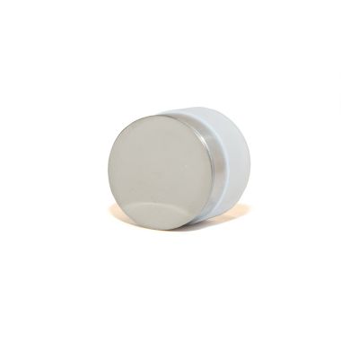
LUXOR Sample Holder for Mounted/Embedded Samples
Product Code:
73724
Manufacturer: LUXOR
The LUXOR sample holder for mounted/embedded samples can hold all types of samples that are mounted or embedded into resins with diameter ranging from 25 mm to 40 mm and height from 10 mm to 50 mm.
The LUXOR sample holder for mounted/embedded samples can hold all types of samples that are mounted or embedded into resins with diameter ranging from 25 mm to 40 mm and height from 10 mm to 50 mm.
Write Your Own Review
| Photo | Product | Price | |
|---|---|---|---|
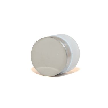
 | LUXOR Gold-Palladium Target | > | |
 | LUXOR Carbon Coater | > | |
 | LUXOR Platinum Coater | > | |
 | LUXOR Gold Coater | > | |
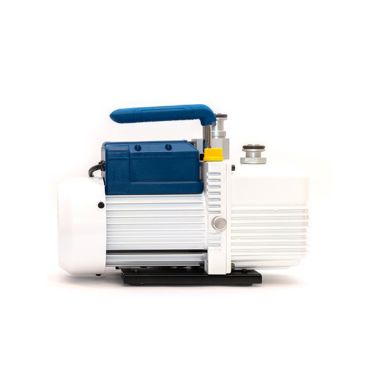 | LUXOR Vacuum Pump | > | |
 | LUXOR Platinum Target | > | |
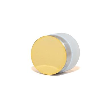 | LUXOR Gold Target | > |