

Sample preparation in Electron Microscopy is one of the most important factors in obtaining high quality data.
A key phrase to remember is if you put “rubbish in” you’ll get “rubbish out”.
The main characteristic of high-quality TEM specimens, is that they are very thin. Ideally, a thickness that is close to the mean free path of the electrons that travel through the samples. For instance, High Resolution Imaging will need samples with a thickness of 10nm. For electron Energy Loss Spectroscopy (EELS) between 10 to 50nm and for diffraction contrast around 300 to 500 nanometres.
But how do you achieve the appropriate specimen thickness?
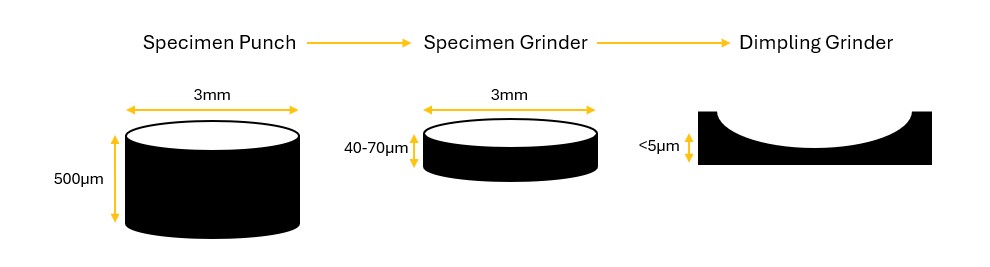
A sample will normally start as a Bulk material. Using the Fischione Model 130 specimen punch a 3mm disk can be sectioned from the bulk material. This disk will be in the region of 500μm or thicker. Thus, the size needs to be reduced further. This can be achieved with the use of the Fischione Model 160 specimen grinder. The grinder can reduce the thickness to roughly 20μm. Then by utilising a high precision Fischione Model 200 dimple grinder the thickness can be reduced to less than 5μm.
The final step to prepare an ideal sample for TEM analysis is Ion Milling. Once the sample thickness is approximately 5μm or less, the Fischione 1051 TEM Ion Mill can be used to reduce the thickness of the specimen further, potentially until it perforates.
During this procedure the user needs to observe the specimen closely and wait for the coloured fringes to appear. For example, when silicon is very thin (<1µm), changes in the colour of its fringes will directly correlate with changes in the specimen thickness. By milling at higher kV until coloured fringes are first observed and then milling at lower kV until perforation, one can accurately endpoint the milling process.
After then being plasma cleaned, ideally using the Fischione 1070, the specimen will be ready for analysis within the microscope.
This article covers the basic principles required to achieve electron transparency, but not all samples are considered equal. Please visit our website or contact us for more in-depth preparation steps on 01582 764334 or click here to email.
Lambda Photometrics is a leading UK Distributor of Characterisation, Measurement and Analysis solutions with particular expertise in Electronic/Scientific and Analytical Instrumentation, Laser and Light based products, Optics, Electro-optic Testing, Spectroscopy, Machine Vision, Optical Metrology, Fibre Optics and Microscopy.
-

Fischione Model 130 Specimen Punch
- A precision ground punch and die plate eliminate specimen stress and distortion
- A spring-loaded return plunger keeps the disk specimen on the die plate surface for convenient handling
- Available in standard sizes of 1 mm, 2.3 mm, and 3.0 mm
- Other sizes available upon request
-

-
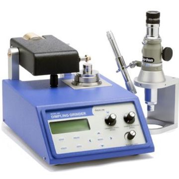
Fischione Model 200 Dimpling Grinder
- Controlled thinning rate
- Precise
- Easy to use
- Automated operation
- Alignment microscope
-
Fischione Model 1051 TEM Mill (Ion Milling)
- Ion milling for TEM
- High energy operation for rapid milling; low energy operation for specimen polishing
- Two independently adjustable TrueFocus ion sources
- Ion source maintains its small beam diameter over a wide range of operating energies (100 eV to 10 keV)
- Continuously adjustable milling angle range of -15 to +10°
- Adjustable 10-inch touch screen with a user-friendly interface for simple setup of milling parameters
- Specimen holder with x-y adjustment
- In situ viewing and image capture during milling
- Liquid nitrogen-cooled specimen stage
-

Fischione Model 1070 NanoClean
- Simultaneously cleans specimens, specimen holders, and specimen mounts
- No change to elemental composition or structural characteristics
- Multiple gas inlets with mixing capabilities
- Evacuates vacuum storage containers